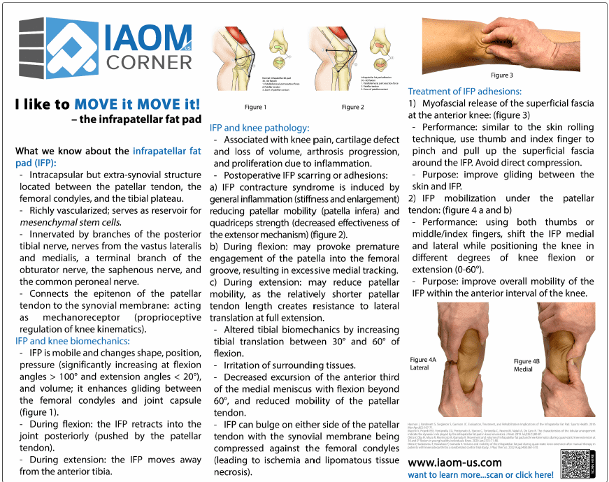
What we know about the infrapatellar fat pad (IFP):
- lntracapsular but extra-synovial structure located between the patellar tendon, the femoral condyles, and the tibial plateau.
- Richly vascularized; serves as reservoir for mesenchymal stem cells.
- Innervated by branches of the posterior tibial nerve, nerves from the vastus lateralis and medialis, a terminal branch of the obturator nerve, the saphenous nerve, and the common peroneal nerve.
- Connects the epitenon of the patellar tendon to the synovial membrane: acting as mechanoreceptor (proprioceptive regulation of knee kinematics).
IFP and knee biomechanics: - IFP is mobile and changes shape, position, pressure (significantly increasing at flexion angles > 100° and extension angles < 20°), and volume; it enhances gliding between the femoral condyles and joint capsule (figure 1).
- During flexion: the IFP retracts into the joint posteriorly (pushed by the patellar tendon).
- During extension: the IFP moves away from the anterior tibia.

