Gutteck N, Schilde S, Delank KS. Dtsch Arztebl Int 2019; 116: 83-8.
Abstracted by Kasey Miller PT, DPT, COMT Kansas City, Missouri – Fellowship Candidate, IAOM-US Fellowship Program & Jean-Michel Brismée, PT, ScD, Fellowship Director, IAOM-US Fellowship program
Research: The purpose of this review article was to present the clinical findings of the pathologies that most commonly cause plantar foot pain and an overview of treatment options.
Methods: The authors carried out a selective survey of literature in the PubMed database. The search terms they included were “plantar fasciitis,” “plantar spur,” “Morton neuroma,” “metatarsalgia,” “transfer metatarsalgia,” “Freiberg’s infraction,” and “heel pain.” After they collected the articles from their search, they took into consideration national and international recommendations about treatment of these pathologies and the clinical presentation of each pain generator. They took special interest in plantar fasciitis and metatarsalgia. While looking into these two pathologies, they specifically wanted to summarize the following categories: symptoms and clinical picture, epidemiology, anatomy and biomechanics, pathogenesis and risk factors, clinical examination, diagnostic imaging, and treatment.
Results: According to previous publications, plantar fasciitis is multifactorial in origin and is viewed as a mechanical overloading reaction to multiple instances of microtrauma. The risk factors are thought to be shortening of the calf muscles, overweight, long periods of employment in active occupations, and deformities of the foot. They found plantar fasciitis to be most common between 45 and 65 years of age. Symptoms that begin to indicate the plantar fascia, as the primary pain generator is medial plantar pain and pain during the first few steps after getting out of bed in the morning or following periods of inactivity. Along with the patient’s subjective examination, a thorough clinical examination is most reliable to detect plantar fasciitis.1 The review references the Silvferskjöld test as a helpful test to determine presence of shortened calf muscle, causing increased stress on the plantar fascia (Figure 1). It was also found that the pain can be provoked by palpation of the area of insertion of the plantar fascia to the calcaneal tubercle and intensified by forced extension of the lesser toes. Although plantar fasciitis is very common, it was found that 90% of patients could have pain relief with conservative treatment within 6 months. The primary conservative treatment is a stretching program of the calf muscles and the plantar fascia. Digiovanni et al. investigated the long-term outcomes of a standardized plantar fascia stretching program, which reported freedom from pain in 92% of patients after 2 years’ follow up (Figure 2). Secondary treatment options include specific foot intrinsic exercises, eccentric exercising, weight loss, or fascia training of the ankle joint.2 Factors that lead to chronic overload and exertion of tension on the plantar fascia should always be considered when deciding how to treat plantar fasciitis. Lee et al. found the use of insoles to relieve the medial segment of the longitudinal arch of the foot is associated with significant pain reduction and functional improvement. If conservative treatment has failed, surgical treatment is an option. However, it should be noted that in 90-95% of patients, combined conservative treatment measures achieve adequate pain relief within 12 months.3
In metatarsalgia, it is helpful to divide the symptom complex into primary and secondary metatarsalgia. Primary metatarsalgia comprises pain of mechanical origin. Pain caused by underlying diseases (Morton neuroma, Kohler disease II, rheumatoid arthritis) is secondary metatarsalgia. Due to the diversity of pain generators that can be at the root of metatarsalgia one has to assume the etiology of metatarsalgia is multifactorial. With the numerous causes of metatarsalgia, imaging is commonly very helpful. MRI is valuable for tumor diagnosis, for evaluation of ligaments, tendons, and plantar plate, and for detection of Morton neuroma. Pedobarography is useful to determine the pressure relationships in the foot and for monitoring in the wake of treatment or surgical correction. Lastly, interdigital neuroma, bursitis, ganglion, joint effusion, and tendon pathology are all visualized well by sonography. Conservative treatment includes the use of insoles or specially adapted shoes to modify the pressure distribution and improve the joint axes. Raising the second and third metatarsal heads by means of an individually molded insole with a retrocapital cushion or a so-called butterfly roll relieves the pressure in up to 60% of patients.4


Figure 1: The Silvferskjöld test is used to determine the presence of a shortened calf muscle. To ensure movement is coming from the talonavicular joint, maintain subtalar neutral throughout. First, flex the knee to reduce tension on the gastrocnemius and evaluate tension on the soleus by assessing the ankles ability to dorsiflex (Left). Next, extend the knee to tighten the gastrocnemius and then assess the ankles ability to dorsiflex (Right). This gives you information about the tightness of the gastrocnemius.


Figure 2: Digiovanni et al compared plantar fascia specific stretching (left) to that of traditional achilles tendon stretching (right) in those with chronic plantar fasciitis. The stretches were performed at 10 reps for 10 seconds 3 times a day. Both groups showed marked improvement two years after with an especially high rate for the plantar fascia-stretching program.5
IAOM-US Comments: Those with medial plantar foot pain as reported in this article can be indicative of plantar fasciitis, but the IAOM-US recognizes that there are other structures that can cause this pain that may be mistaken for plantar fasciitis. Clinically, the three most common pain generators of heel pain are plantar fasciitis, medial plantar nerve irritation (distal tarsal tunnel syndrome), and flexor digitorum brevis (FDB) tendopathy6. And it is vital that we differentiate each one of these, as the treatment for each is completely different.
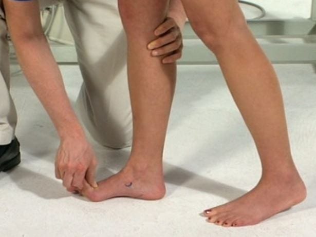
As discussed in the review article, plantar fasciitis will present with plantar-medial heel pain with increased pain with weight bearing after periods of inactivity or first thing in the morning. Pain decreases with activity but with continued activity, the pain begins to intensify. The IAOM-US advocates for a dynamic supination test to help differential diagnosis (Figure 3) as well dysfunction identification.
Distal tarsal tunnel syndrome, also known as jogger’s foot, involves medial plantar nerve irritation at abductor hallucis. This is often confused with plantar fasciitis due to the location of the pain. Some ways to differentiate medial plantar nerve irritation from plantar fasciitis include the presence of burning, tingling or cramping as well as sensory loss indicating nerve irritation. The medial plantar distribution includes the plantar surface of the medial 3 toes, with less commonly foot intrinsic wasting in severe cases. One of the most prominent ways to distinguish medial plantar nerve irritation from plantar fasciitis is a positive neural tension test (Figure 4). Patient history is another important aspect of the clinical examination that helps identify medial plantar nerve irritation. The patient will report painful gait, increased pain with increased activity throughout the day, and decreased pain in the morning. Lastly, during palpation, the tender spot is more medial on distal calcaneal tuberosity as opposed to directly on the calcaneal tuberosity with plantar fasciitis.
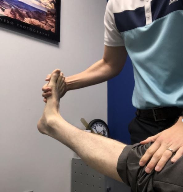
Flexor digitorum brevis (FDB) tendopathy is another pathology that can present with medial heel pain and can be misdiagnosed as plantar fasciitis. This pathology is commonly seen in long distance runners. Using selective tissue testing, the most painful test for FDB should be resisted testing of the toes, which helps distinguish between medial plantar nerve irritation and plantar fasciitis. Another way to differentiate between fasciitis and FDB is to perform palpation of the insertion with the plantar fascia on slack and compare with palpation while plantar fascia is taut with toes passively extended. FDB tendopathy will be most painful to palpation while the plantar fascia is on slack due to the fact that when the fascia is tightened, there is a bridge that is created over the FDB insertion so palpation of the muscle is very difficult and less or not painful (Figure 5).
An important point to make involves the use of palpation. While this article utilizes palpation as a sole test for the diagnosis of plantar fasciitis, the IAOM-US suggests using palpation as a confirmation of your clinical examination. No one test should stand-alone. Due to the proximity of the plantar fascia, medial plantar nerve, and flexor digitorum brevis, using palpation as a single diagnostic test sets the clinician up for failure.
Once the proper cause of heel pain has been identified, implementing the optimal treatment will be much clearer. As previously mentioned, if a patient has plantar fasciitis a proper stretching program of the heel-cord should be initiated. It has also been found that shoe inserts, steroid injection, and custom made night splints can be beneficial for those with plantar fasciitis.7 The use of foot intrinsic strengthening and manual therapy such as soft tissue and joint specific mobilizations have also been found to be beneficial.8 In regards to the treatment of medial plantar nerve irritation, the use of a posterior heel wedge or full contact orthotic without an arch can alleviate pressure to reduce the acute inflammation of the nerve. Once the intensity of pain has reduced, implementing neural mobilization is greatly beneficial (Figure 6). And lastly, the treatment of FDB tendopathy should include transverse friction of the origin on the medial calcaneal tubercle. In order to effectively perform transverse friction, the plantar fascia should be on slack by flexing the toes while the ankle is plantar-flexed or in neutral. It is also important to recognize which stage the tendopathy is in as reducing mileage, implement pool running or instruct the patient in cycling may be beneficial until the pain lessens.
Differentiating structures that cause heel pain can be very difficult. And as previous discussed, it is often only one or two special tests that can improve the clinician confidence in the diagnosis and start the optimal treatment approach. However, if one fails to correctly identify the source of heel pain, the patient’s progress can be slower.

Figure 3: Dynamic supination test is started with the patient shifting their weight onto the forward foot. During passive extension of the great toe, elevation of the medial arch and lateral rotation of the tibia should occur. If the arch fails to rise, there is dysfunction in windlass mechanism of plantar aponeurosis. Reproduction of patient’s heel pain with passive extension of the great toe is most often indicative of plantar fasciitis. When combined with extension of all MTP joints, this may also increase strain in plantar nerve, requiring further differential diagnosis between plantar fasciitis and medial plantar nerve irritation.

Figure 4: Medial plantar nerve can be emphasized during a straight leg raise to assess the sensitivity of the nerve. While performing the straight leg raise neural tension test, passively extend the medial three toes and add dorsiflexion of the talocrural joint along with eversion. If this reproduces their medial heel pain, this indicates involvement of the distal tarsal tunnel.


Figure 5: Differential test between fasciitis and FDB includes palpation of its insertion while plantar fascia is on slack (left) and when the plantar fascia is taut (right). Plantar fasciitis will be most painful while the fascia is taut while FDB tendopathy will be most past painful while the plantar fascia is on slack.
References:
- Alazzawi S, Sukeik M, King D, Vemulapalli K: Foot and ankle history and clinical examination: a guide to everyday practice. World J Orthop 2017; 8: 21-9.
- Rano JA, Fallat LM, Savoy-Moore RT: Correlation of heel pain with body mass index and other characteristics of heel pain. J Foot Ankle Surg 2001; 40: 351-6.
- Kinley S, Frascone S, Calderone D, Wertheimer SJ, Squire MA, Wiseman FA: Endoscopic plantar fasciotomy versus traditional heel spur surgery; a prospective study. J Foot Ankle Surg 2993; 32: 595-603.
- Holmes GB, Timmerman L: A quantitative assessment of the effect of metatarsal pads on plantar pressures. Foot Ankle 1990; 11:141-5.
- DiGiovanni B et al. Plantar Fascia-specific stretching exercise improves outcomes in patients with chronic plantar fasciitis: A prospective clinical trial with two-year follow-up. J Bone Joint Surg Am 2006: 1775-1781.
- Thomas J, et al. The Diagnosis and Treatment of Heel Pain: A Clinical Practice Guideline-Revision. The Journal of Foot and Ankle Surgery. 2010; 49 (2010): 1-19.
- Cole C, Seto C, Gazewood, J. Plantar fasciitis: evidence-based review of diagnosis and therapy. Am Fam Physician. 2005; 72(11): 2237-42.
- Cleland JA, et al. Manual physical therapy and exercise versus electrophysical agents and exercise in the management of plantar heel pain: a multicenter randomized clinical trial. J Orthop Sports Phys Ther. 2009; 39(8):573-85.
